


Dental Exams and X-Rays Explained

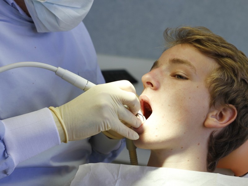
Dental exams and X-rays are important tools dentists use to understand the complete condition of your oral health. A dental exam focuses on evaluating the visible structures of the mouth, including the teeth, gums, bite, tongue, and other soft tissues. This evaluation helps identify early signs of common dental concerns such as tooth decay, gum inflammation, enamel wear, or changes in the way the teeth come together when biting.
Dental X-rays, also called dental radiographs, provide images of areas that cannot be seen during a visual exam. These images allow dentists to view the internal structure of the teeth, the roots, surrounding bone, and spaces between teeth. Because many dental problems begin below the surface, X-rays help detect conditions such as cavities between teeth, infections near the roots, bone loss, impacted teeth, or abnormalities in the jaw. Together, dental exams and X-rays give dentists a complete picture of both the visible and hidden aspects of oral health. This comprehensive evaluation helps monitor changes in the teeth and jaw over time and allows dentists to identify potential concerns early before they become more serious.